2. 深圳市第二人民医院脊柱外科, 广东 深圳 518035 ;
3. 深圳市龙华新区中心医院耳鼻咽喉头颈外科, 广东 深圳 518110
2. Department of Spine Surgery, Shenzhen Second People's Hospital Clinical College, Shenzhen 518035, Guangdong, China ;
3. Department of Otolaryngology & Head and Neck Surgery, Central Hospital of Longhua New District, Shenzhen 518110, Guangdong, China
内镜经口寰枢椎手术因其入路简单,无需牵拉重要血管、神经等优点,深受广大临床医生的喜爱。2015年3月至2015年10月我们对5例新鲜尸头标本采用内镜经口入路手术模拟以及寰枢椎CT测量,以期得到寰枢椎详细解剖数据,观察咽后、椎前、颈椎椎体结构层次以及重要血管神经的相互毗邻关系,明确内镜经口手术可显示及操作范围。探索内镜经口入路的恒定解剖标志,并探讨内镜经口寰枢椎手术的可行性及先进性。
1 资料与方法 1.1 标本材料与设备新鲜尸头5例,其中经颈总动脉行红色乳胶灌注1例,未分性别,无籍贯及年龄记录。所有尸头均由深圳大学医学院人体解剖教研室及深圳市红十字会遗体捐献中心提供。
内镜系统: 使用4mm的0°、30°硬性鼻内镜、冷光源系统、高清摄像头、高清工作站(Xion公司,德国)。
1.2 内镜经口寰枢椎手术操作步骤①于“C”形头架上固定尸头标本,开口器打开口腔,牵拉软腭,暴露视野;②内镜下扪及寰椎前结节,以寰椎前结节为中心,纵行切开咽后壁黏膜、椎前筋膜,离断、剥离头长肌、颈长肌,分离寰枢椎关节囊及韧带,暴露寰枢椎腹侧骨性结构;③磨除寰椎前弓,暴露齿突,离断齿突基底部,分离齿突附着韧带,切除齿突,显露寰椎十字韧带,切开十字韧带,显露后方覆膜及硬脊膜;④移动内镜,显露两侧侧块关节,切开侧块关节囊,向后外侧分离,暴露位于侧块关节外侧的椎动脉。
1.3 寰枢椎CT测量选取临床患者20套正常CT片,确定可准确测量的指标包括:寰椎前弓长度、寰椎前结节厚度、寰椎侧块横径、寰椎侧块矢状径、寰椎两侧横突孔内侧间距、寰椎两侧横突孔外侧间距、齿突后倾角、枢椎两侧横突孔内侧间距、枢椎两侧横突孔外侧间距。CT采用薄层扫描(0.75mm),在CT机上直接读数(如图 1所示)。

|
图 1 CT测量 Figure 1 Results of CT measurement |
采用SPSS 19.0统计软件包,测量结果以x±s表述,并对左右侧对应数据进行配对设计的t检验。检验水准α=0.05,P<0.05为差异有统计学意义。
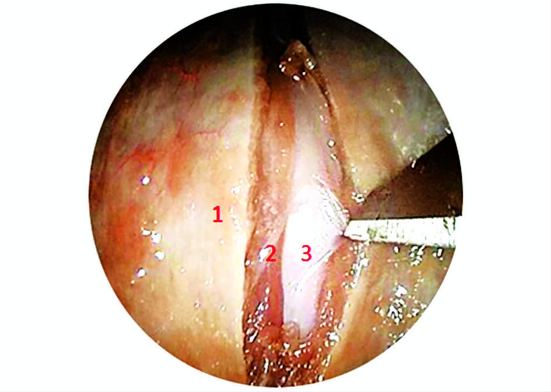
2 结 果 2.1 内镜观察结果 2.1.1 椎前软组织咽后壁[1-2]由内至外分为黏膜层、纤维层、肌肉层、外膜层4层,切开分离过程中,可见由纤维层于中线处形成咽缝,为咽缩肌附着处。其后为咽后间隙,分布着疏松结缔组织及小静脉丛(图 2)。钝性分离至椎前筋膜,切开筋膜,暴露椎前间隙,可见其内头长肌、颈长肌以及寰枕、寰枢关节囊及韧带。离断肌肉、韧带,切开关节囊,向两侧分离,暴露寰枢椎腹侧骨性结构。

|
图 2 内镜经口咽后壁黏膜切开 Figure 2 Endoscopic view of midline vertical mucosal incision over the posterior pharyngeal wall |
内镜下可见寰椎[3-5]前弓(图 3)及两侧侧块,前弓正中隆起,称之为寰椎前结节(图 3),是内镜经口寰枢椎手术的重要定位标志。前弓两侧连接侧块,侧块分别与枕髁及枢椎侧块构成寰枕、寰枢关节,寰枢椎侧块关节是寰枢椎前路固定手术中常用螺钉固定位点。磨除前弓后可见其后方齿突,前弓后方凹陷,与齿突相关节,称之齿突凹。

|
图 3 内镜下寰枢椎腹侧 Figure 3 Close-up view showing the entire anterior tubercle of atlas and part of C1 and C2 body |
内镜下可见枢椎椎体向上伸出,形成齿突[4-6],原为寰椎椎体,生长发育中脱离寰椎,与枢椎融合形成,是上颈椎运动的枢纽。齿突(图 4),前后分别与寰椎齿突凹及寰椎横韧带形成关节,限制其前后运动;两侧及尖部通过翼状韧带及齿突尖韧带与枕骨相连,限制其两侧及旋转运动。齿突与枢椎椎体附着处骨质薄弱,易发生骨折,是影响寰枢椎稳定性的重要因素之一,一般可前路由椎体向上置入螺钉,行复位固定。

|
图 4 内镜下前弓磨除后暴露齿突和枢椎体 Figure 4 Close-up view showing the entire odontion process with part of C2 body |
摘除齿突后,可见坚韧、厚实的寰椎横韧带(图 5),两侧连接侧块,上下通过纤维索连接枕骨及枢椎,统称寰椎十字韧带。切开韧带,依次分离,可见覆膜、硬脊膜及脊髓等(图 6)。向两侧移动内镜可见两侧侧块关节面[7],剥离关节囊及表面附着韧带,见侧块宽大、厚实,骨质坚硬,适合复位固定手术中钢板及螺钉的固定。

|
图 5 寰椎横韧带暴露 Figure 5 Close-up view showing thetransverse ligament of atlas |
沿侧块向外后分离,可见走行于横突孔中的椎动脉[8](图 7),解剖学中分为4段,与寰枢椎相关的为第3段,即椎动脉寰椎部。此段由枢椎横突孔向上弯曲走行,穿过寰椎横突孔,向后走行于寰椎椎动脉沟内,转而向内上,进入硬膜。

|
图 6 切开十字韧带后暴露覆膜及硬脊膜 Figure 6 Close-up view showing the spinal dura mater and tectorial membrane |

|
图 7 内镜下寰枢侧块关节和椎动脉(右) Figure 7 Close-up view showing the vertebral artery and lateral mass of atlas and axis (right) |
寰枢椎CT测量数据,见表 1。
| 表 1 寰枢椎CT测量数据 Table 1 The results of CT measurement of Atlanto-axial vertebral |
对寰枢椎左右对称的测量数据进行配对t检验,结果显示P值均>0.05,统计学上无显著性差异,见表 2。
| 表 2 寰椎侧块双侧测量结果对比(n=20, x±s, mm) Table 2 The comparison of data on both sides of lateral mass of atlas (n=20, x±s, mm) |
经口入路常用于处理寰枢椎腹侧病变,由于入路狭小、深在,毗邻延脊髓、椎动脉等重要的神经和血管结构,且经口入路为相对污染区,有一定发生感染的风险,使其发展受限。但随着内镜技术兴起、发展以及引入,使得在小切口的基础上,也可以获得满意的视野范围,清晰的辨认各解剖结构的关系,保证操作的精确性。
3.1 内镜经口寰枢椎手术可行性根据内镜解剖观察,配合广角内镜,术中暴露范围向上可达中下斜坡,向下最多可至第三颈椎上部,显露的总宽度达(40.1±5.2)mm,寰椎前弓、侧块、齿突及枢椎椎体都可以清晰显露[9-10]。对于寰枢椎腹侧占位性病变,如:结核病灶、良性肿瘤以及经过选择的局限性恶性肿瘤,都可以直视下予以完整或分块切除,亦可内镜协助下完成植骨、钢板固定。
通过对寰枢椎CT测量数据的分析以及内镜解剖观察,可得出一手术安全区域。椎动脉走行于寰枢侧块关节外侧,为避免损伤椎动脉,可界定两侧的安全边界:上方为寰枢侧块关节外缘,下方为枢椎体外缘。内镜下上方常规可显露至寰椎前弓上缘或斜坡下部,下方可显露至C2/3椎间盘或C3椎体上部。
3.2 内镜经口寰枢椎手术注意事项咽后壁软组织分离中,界定两侧安全边界后,直接用电刀、电凝分离。分离软组织后,可根据手术要求,以寰椎前结节为中心,磨除寰椎前弓[11]。齿突切除[12]可根据手术需要,由齿突尖向下帽状磨除,或先离断基底部,小心分离后,完整切除。内镜解剖观察寰椎横韧带中线较厚、且距脊髓较近,两侧韧带较薄、距后方脊髓较远,若行寰椎横韧带切除,建议在侧块附着处离断。椎动脉走行于寰枢侧块关节囊外侧,在行侧块关节打磨手术时,应注意关节囊内操作,避免突破关节囊、损伤椎动脉。
3.3 内镜经口Ⅰ期行复位内固定可行性内镜经口寰枢椎术后,常因骨质缺损以及关节囊、韧带、肌肉结构破坏,导致术后出现寰枢椎不稳等并发症,因此,内镜协助下Ⅰ期完成植骨、钢板固定显得尤为重要。常用的寰枢椎钢板系统[15]有:Harms钢板、寰枢关节锁定钢板、寰枢椎复位钢板系统、重建钢板。Kandziora等[16]等对寰枢椎标本进行测量,得出寰椎螺钉进钉位置的形状与侧块前表面相似,而枢椎螺钉进钉位置的形状呈“ V”字形。
3.4 内镜经口寰枢椎手术先进性传统手术为了获得满意手术视野,需要长时间维持大程度张口,有时需切开软腭、硬腭,甚至配合行上颌骨、下颌骨截断等。手术创伤大,术后并发症多,使得经口入路难以临床上广泛展开。 相对于传统手术方式来说,内镜经口寰枢椎手术有着无可取代的优越性。利用高清内镜显示系统,我们可以将狭小、深在的术腔放大到高清显示屏上,有利于术者观察手术过程,辨析周围解剖关系。通过内镜的移动,配合多角度内镜,还可以清晰观察术野周边情况,可直视下完成前弓切除、齿突摘除、关节打磨,可精确控制切除、打磨的范围、深度,减少对脊髓、椎动脉等重要结构的损伤,减少术中损伤,降低术后并发症发生率。 但内镜经口寰枢椎手术仍存在不足:内镜显示是二维图像,缺乏三维立体结构感,手术过程中对距离的远近把握不清;内镜下术腔狭小,需借助各种器械,无法直接操作;相应的配套实施不完善。
| [1] |
Hsu W, Wolinsky J P, Gokaslan Z L, et al. Transoral approaches to the cervical spine[J].
Neurosurgery,2010, 66 (3) : 119-125.
( 0) 0)
|
| [2] |
黄选兆.
实用耳鼻咽喉头颈外科学[M]. 北京: 人民卫生出版社, 2008 : 288 -289.
( 0) 0)
|
| [3] |
Doherty B J, Heggeness M H. The quantitative anatomy of the atlas[J].
Spine,1994, 19 (22) : 2497-500.
( 0) 0)
|
| [4] |
柏树令.
系统解剖学[M]. 北京: 人民卫生出版社, 2009 : 17 -46.
( 0) 0)
|
| [5] |
胥少汀.
实用骨科学[M]. 北京: 人民卫生出版社, 2012 : 1935 -1969.
( 0) 0)
|
| [6] |
Doherty B J, Heggeness M H. Quantitative anatomy of the second cervical vertebra[J].
Spine,1976, 20 (5) : 513-517.
( 0) 0)
|
| [7] |
Li Shi, Chai Yu, Li Yikai, et al. Quantitative anatomy of the tubercle of lateral mass of atlas and dens of axis and its clinical significance[J].
Chin J Anat,2014, 37 (2) : 212-216.
( 0) 0)
|
| [8] |
Fine A D, Cardoso A, Rhoton A L Jr. Microsurgical anatomy of the extracranial-extradural origin of the posterior inferior cerebellar artery[J].
J Neurosurg,1999, 91 (4) : 645-652.
( 0) 0)
|
| [9] |
Wang Z, Xia H, Wu Z, et al. Detailed anatomy for the transoral approach to the craniovertebral junction: an exposure and safety study[J].
J Neurol Surg B Skull Base,2014, 75 (2) : 133-139.
( 0) 0)
|
| [10] |
Wang Y, Yang M, Zhang H, et al. Exploring the Safety Range via the Transoral Approach to the Craniovertebral Junction[J].
J Craniofac Surg,2014, 25 (4) : 1473-1475.
( 0) 0)
|
| [11] |
Urculo-Bareo E, Alfaro-Baca R, Astudillo-Alarcón E, et al. Transoral decompression of the brain stem in a basilar impression secondary to rheumatoid arthritis. Description of a case and survey of the literature[J].
Rev Neurol,2003, 36 (9) : 846-851.
( 0) 0)
|
| [12] |
Tanriverdi O, Tugcu B, Gunaldi O, et al. The selective odontoidectomy: endoscopic endonasal approach to the craniocervical junction[J].
J Craniofac Surg,2014, 25 (4) : 1482-1487.
( 0) 0)
|
| [13] |
Song K J, Lee K B, Kim K N, et al. Treatment of odontoid fractures with single anterior screw fixation[J].
J Clin Neurosci,2007, 14 (9) : 824-830.
( 0) 0)
|
| [14] |
Ma W, Xu N, Hu Y, et al. Unstable atlas fracture treatment by anterior plate C1-ring osteosynthesis using a transoral approach[J].
Eur Spine J,2013, 22 (10) : 2232-2239.
( 0) 0)
|
| [15] |
Ai F, Yin Q, Wang Z, et al. Applied anatomy of transoral atlantoaxial reduction plate internal fixation[J].
Spine,2006, 31 (2) : 128-132.
( 0) 0)
|
| [16] |
Kandziora F, Schulze-Stahl N, Khodadadyan-Klostermann C, et al. Screw placement in transoral atlantoaxial plate systems: an anatomical study[J].
J Neurosurg,2001, 95 (1) : 80-87.
( 0) 0)
|
 2016, Vol. 30
